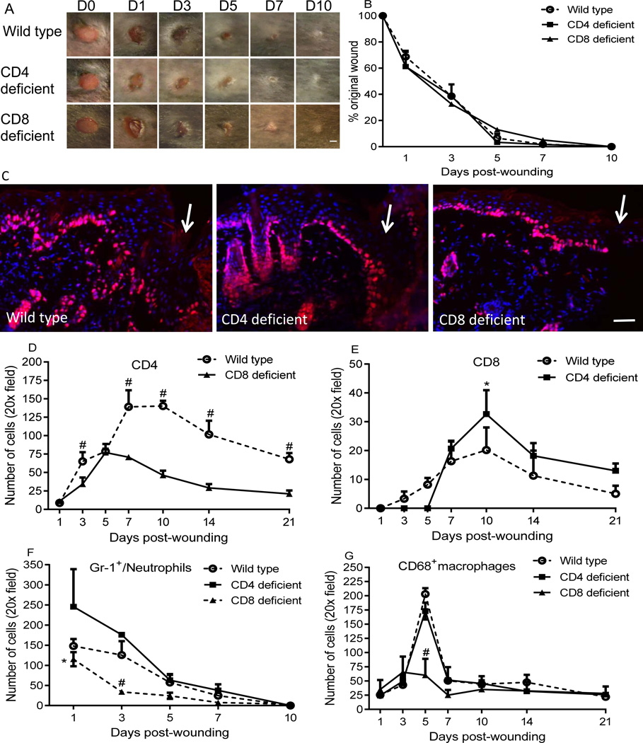
Figure 2.
CD4 or CD8 lymphocyte deficiency does not impair wound closure, but results in altered CD8+, CD4+, neutrophils and/or macrophage infiltration in the wounds. A) Representative photos of wounds photographed at various time points after injury. B) Percent of the original wound size. N=5 in each group; scale bar=1mm. C) Representative photomicrographs demonstrating immunohistochemical detection of the proliferation marker ki67 at the wound margin at day 3 (days 1 and 5 not shown). Arrows indicate wound edges. Red: ki67, blue: DAPI stained nuclei. Scale bar=100µm. D&E) Time course of numbers of CD4+ and CD8+ cells in the wounds of wild type/CD8 deficient mice, and wild type /CD4 deficient mice respectively. # p<0.001 compared to CD8 deficient mice, * p<0.05 compared to wild type. F&G) Time course of numbers of neutrophils and macrophages. * p<0.05 and # p<0.01 compared to CD4 deficient and wild type mice. Two-way ANOVA followed by a Tukey’s multiple comparisons test was used. Error bars = standard deviation.

