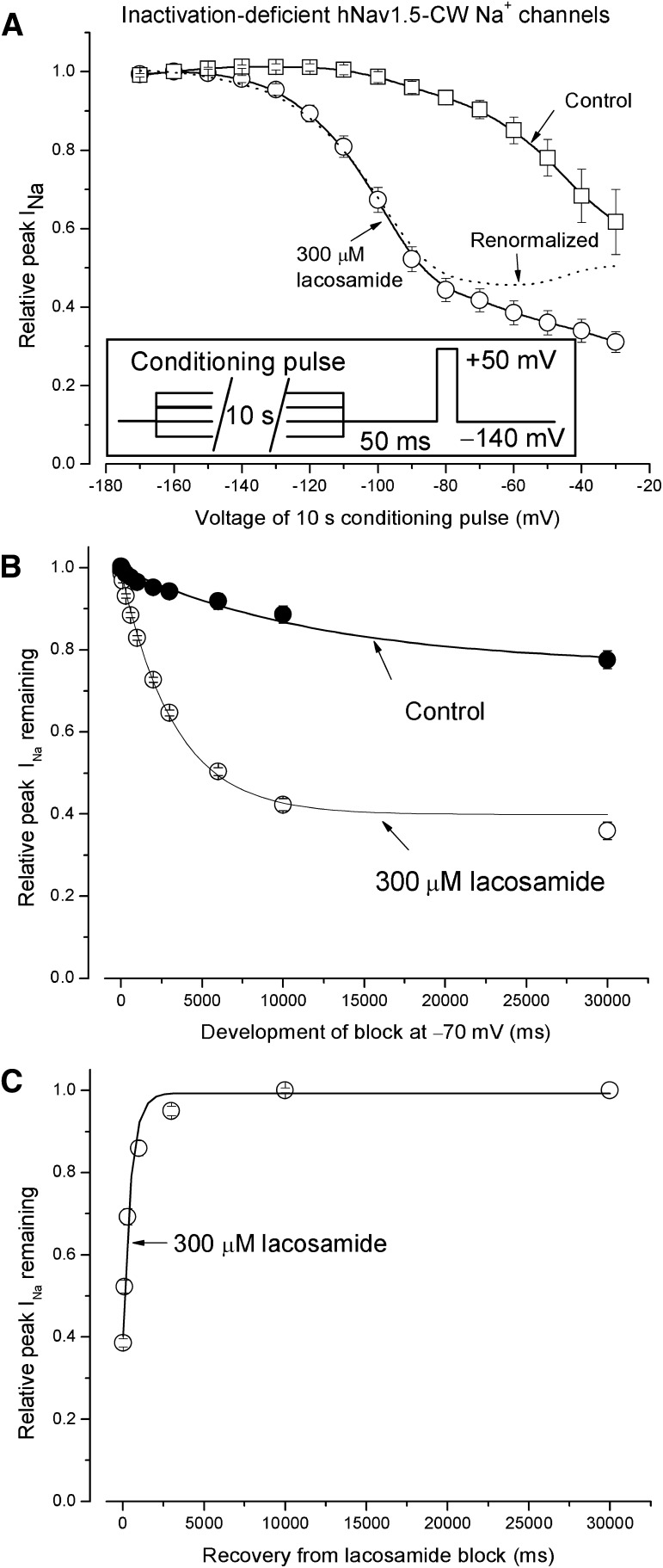
Fig. 6.
Block of inactivation-deficient hNav1.5-CW Na+ channels by lacosamide at various membrane potentials. (A) The voltage-dependent block was measured at various conditioning pulses in the presence (○) or absence of 300 µM lacosamide (□). The inset shows the pulse protocol. Without drug, the peak current amplitude decreased considerably from −100 to −30 mV, probably because of the enhanced slow inactivation in inactivation-deficient hNav1.5-CW mutant Na+ channels. (B) The development of block by 300 µM lacosamide at −70 mV was recorded using hNav1.5-CW Na+ channels and plotted against the time (○). The solid line was the best fitted of the data by a single exponential function with a time constant of 3.28 ± 0.12 seconds (n = 8). Without drug, the time constant was 12.5 ± 3.7 seconds. (C) The recovery from the lacosamide block at −70 mV for 10 seconds was measured at the holding potential −140 mV, with a duration ranging from 0 to 30 seconds before a test pulse (+50 mV for 10 milliseconds) was applied. The relative peak currents were then plotted against the time, and the data were fitted by an exponential function with a time constant of 476 ± 38 milliseconds (n = 8; r2 = 0.99).

