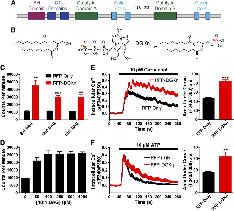
Fig. 1.
Mouse DGKη is catalytically active and enhances GPCR signaling. (A) Domain architecture of DGKη. (B) Schematic of the reaction forming 32P-labeled PA from DAG and radiolabeled ATP. (C) Lysates from HEK293 cells expressing (black) RFP alone or (red) RFP-DGKη were incubated with the indicated DAG substrates, each at 500 µM. (D) 32P-PA production from reactions containing the indicated concentrations of 18:1 DAG, catalyzed by lysates from HEK293 cells expressing RFP-DGKη. (E and F) Calcium mobilization in HEK293 cells expressing (black) RFP alone or (red) RFP-DGKη after stimulation with (E) 10 µM carbachol or (F) 10 µM ATP. AUC measurements extended for 4 minutes from agonist addition. Data in C and D are the average of two experiments performed in duplicate. Data in E and F are the average of three independent experiments; n = 41–104 cells per condition. **P < 0.01; ***P < 0.001 when compared with RFP alone (unpaired t test). All data, including calcium traces, are presented as means ± S.E. C1, PKC homology domain 1; PH, pleckstrin homology.

