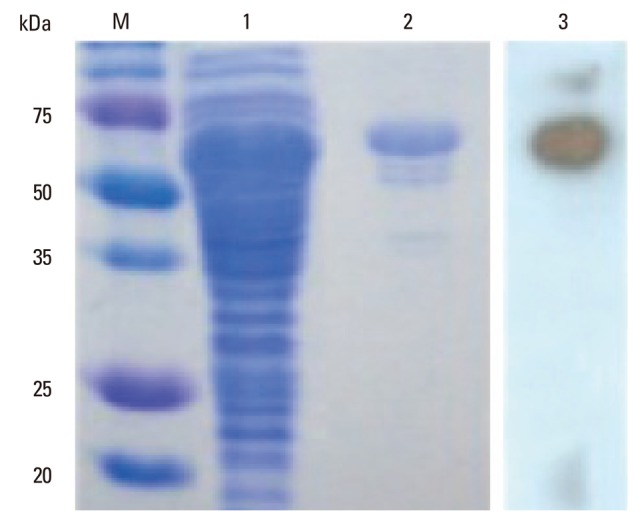
Fig. 1.

Analysis of expression of the MPT64 protein in TB-1 cells. TB-1 cells were inducted with 1 mM IPTG for 4 h, and the cell lysates were analysed by SDS-PAGE, after which the gel was stained with Coomassie blue (lanes 1 to 2) or analysed by Western blot with a mouse polyclonal anti-M. tuberculosis antibody (lane 3). Lane M: pre-stained protein molecular weight markers. Lane 1: cell lysate of TB-1 cells. Lane 2: purified recombinant MPT64. Lane 3: Western blot analysis with anti-MPB64 antibody. TB, tuberculosis; SDS-PAGE, sodium dodecyl sulfate-polyacrylamide gel electrophoresis; IPTG, isopropyl β-D-1-thiogalactopyranoside.
