Figure 2.
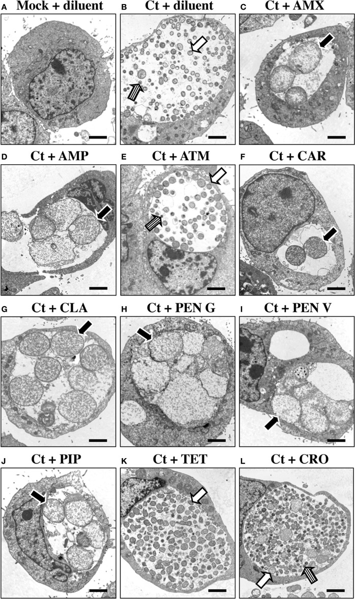
Penicillin-exposure induces chlamydial AB formation. HeLa cells were C. trachomatis-infected and incubated in the absence of antibiotic for 24 h. Infected and uninfected cultures were then refed with medium containing each antibiotic of interest at the 1X concentrations (Table 1). Cells were incubated for an additional 30 h (a total of 54 hpi), fixed and subjected to TEM. (A) Mock-infected cells + ddH2O (diluent). (B) C. trachomatis-infected (Ct) cells + diluent. (C) C. trachomatis + AMX. (D) C. trachomatis + AMP. (E) C. trachomatis + ATM. (F) C. trachomatis + CAR. (G) C. trachomatis + CLA. (H) C. trachomatis + PEN G. (I) C. trachomatis + PEN V. (J) C. trachomatis + PIP. (K) C. trachomatis + TET. (L) C. trachomatis + CRO. Morphologically normal RB and EB are indicated by white and striped arrows respectively. Abberent bodies (AB) are labeled with black arrows. Each photomicrograph is at 7500X magnification; the black bar at the lower right of each panel represent 2 μm.

