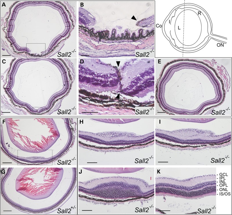
Figure 4.
Histological analysis of eyes dissected from P20 Sall2−/− mice. Top right. Schematic representation of a mouse eye showing the plane of sectioning and approximate anterior location of the observed retinal abnormality (dotted line). Co, cornea; I, iris; L, lens; R, neural retina; ON, optic nerve. (A–E) Coronal sections of the eye from a P20 homozygous Sall2−/− mouse showing a gap in the neural retina visible anteriorly (A and B). In serial sections towards the posterior pole, the tips of the retinal margins meet with residual pigment visible surrounding the tips of the margins (arrowheads in B and D which show magnifications of boxed region in A and C, respectively). (E) More posteriorly, beyond the mid-lenticular region, the retina appears fused and indistinguishable from normal. Note, the lens was removed during processing. (F) Representative coronal section from a homozygous Sall2−/− mouse showing localized elevated lesion of the retina as compared with (G) equivalent coronal section of eye from P20 heterozygous Sall2+/− mouse showing no abnormality. (H–J) Consecutive anterior-to-posterior sections showing magnification of boxed region in (F) showing progressive swelling and disorganization of ganglion cell layer (H), inner plexiform layer (I) and inner and outer nuclear layers (M). In more posterior sections, the retina shows normal lamination (K). GCL, ganglion cell layer; IPL, inner plexiform layer; INL, inner nuclear layer; OPL, outer plexiform layer; ONL, outer nuclear layer; IS/OS, photoreceptor inner and outer segments. Scale bars represent 200 µm in (A, C, F, G and E), 50 µm in (B and D) and 100 µm in (H–K).

