Figure 2.
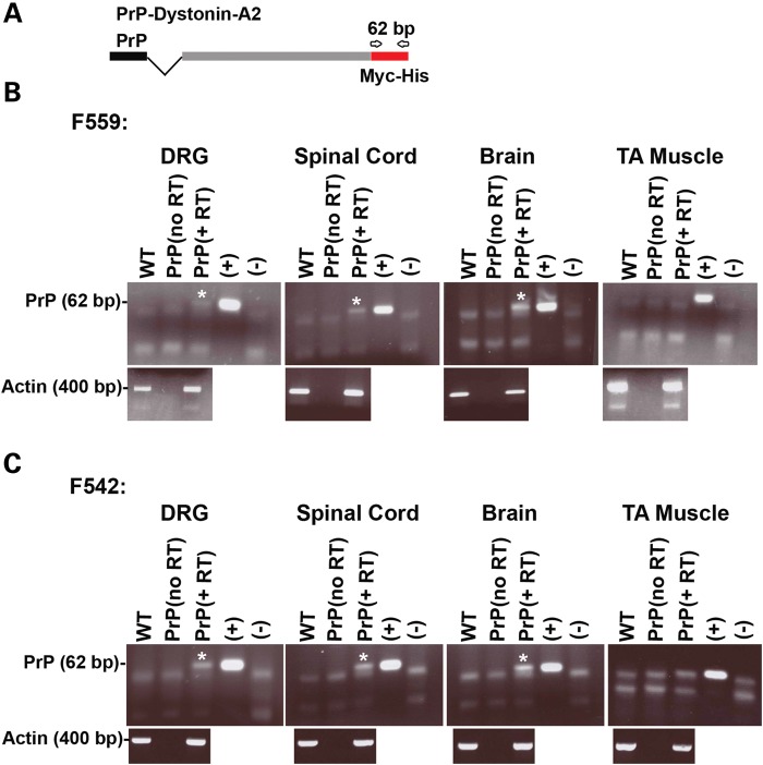
The PrP-dystonin-a2 transgene is expressed in neuronal tissues of transgenic lines 559 and 542. (A) Schematic representation of the location of oligonucleotide primers for the amplification of PrP-dystonin-a2 derived transcripts. Primers amplify a 62 bp fragment that includes the myc/his tag coding sequence. (B and C) RT–PCR analysis of RNA derived from P7 neuronal tissues (DRG, spinal cord and brain) of heterozygous transgenic mice from lines 559 and 542 indicates PrP-dystonin-a2 transgene expression (PrP, +RT lanes). Transgene expression was not evident in tibialis anterior (TA) muscle of either transgenic line. Controls: P7 wild-type (WT) cDNA of neuronal tissues, PrP (no RT, minus reverse transcriptase), (+) and (−) cDNA of F11 sensory neurons transfected or non-transfected with PrP-dystonin-a2 construct, respectively. Actin mRNA amplification served as a positive control for RNA.

