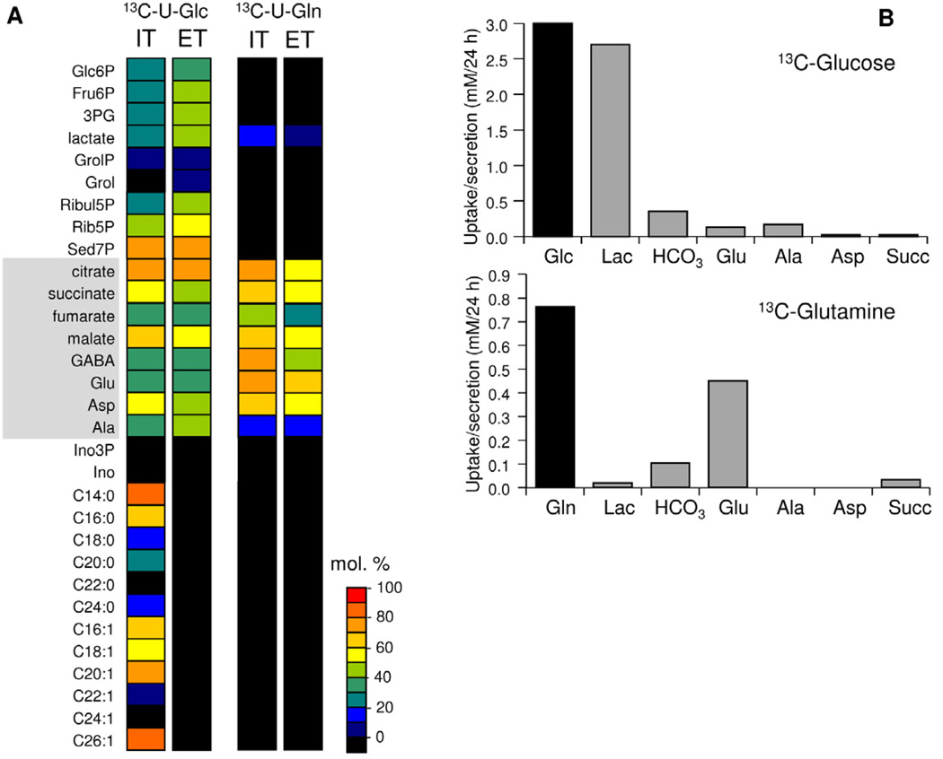
Figure 2. T. gondii tachyzoites catabolize glucose in a complete TCA cycle.
(A) Infected HFF or egressed tachyzoites (ET) were suspended in medium containing either 13C-U-glucose or 13C-U-glutamine for 4 hr. Intracellular tachyzoites (IT) were isolated from host material prior to metabolite extraction. Incorporation of 13C into selected polar metabolites and fatty acids (derived from total lipid extracts) was quantified by GC-MS and levels (mol percent containing one or more 13C carbons) after correction for natural abundance are represented by heat plots. (B) Egressed tachyzoites were incubated in full medium containing either 13C-U-glucose (upper panel) or 13C-U-glutamine (lower panel) in place of naturally labelled glucose or glutamine, respectively. Culture medium was collected at 6, 12 and 24 hr and analysed by 13C-NMR. Rates of utilization of each carbon source are shown in black, while rate of secretion of lactate (Lac), CO2 (detected as H13CO3), glutamate (Glu), alanine (Ala), aspartate (Asp) and succinate (Suc) are shown in grey. See also Figure S2.

