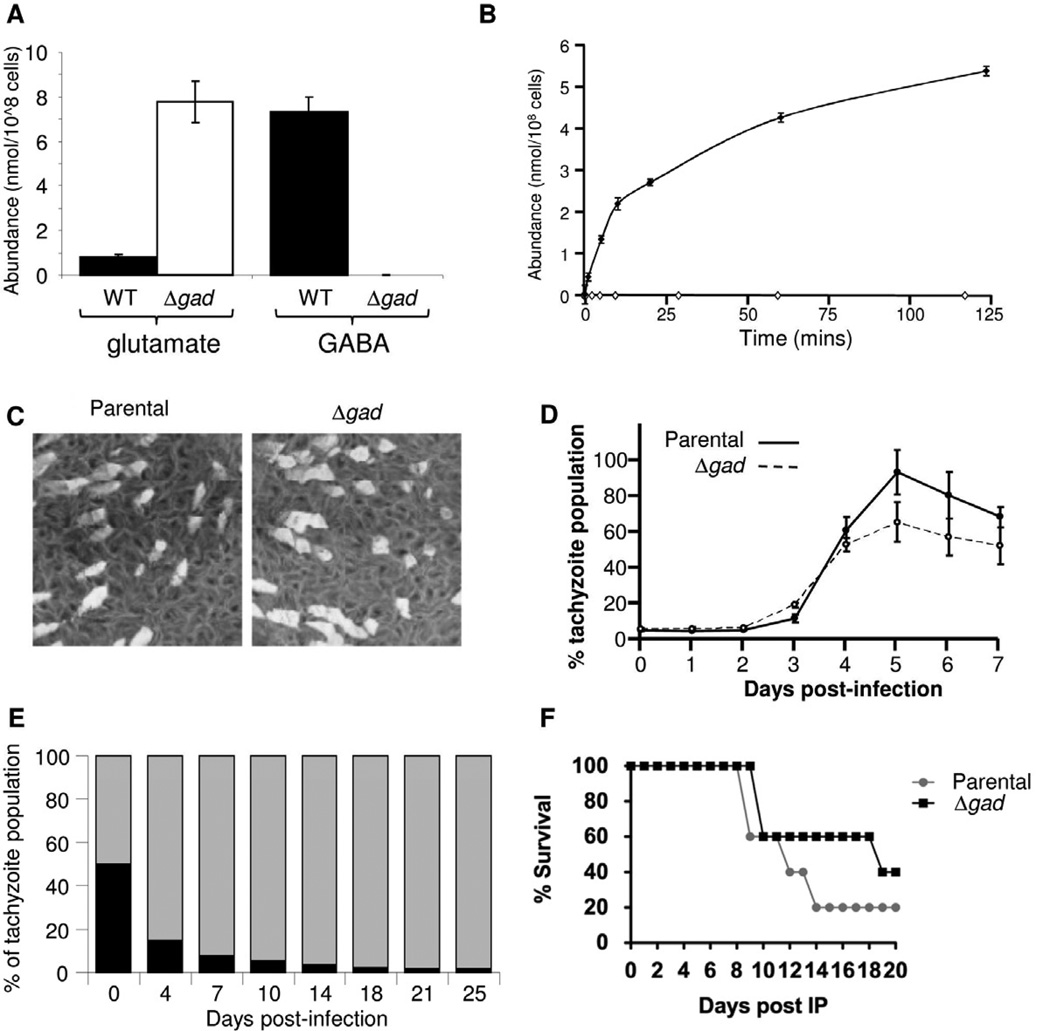
Figure 5. Loss of GAD results in complete ablation of GABA biosynthesis and partial attenuation of parasite propagation.
(A) Intracellular GABA and glutamate levels in parental and Δgad tachyzoites. (B) Cell-free extracts of T. gondii parental (filled circles) and Δgad tachyzoites (open circles) were incubated with 10 mM 13C-U-glutamate in the presence of 1 mM ATP. The synthesis of GABA at indicated time points was measured by GC-MS. Error bars represent standard deviation, where n = 3. (C) Parental and Δgad parasite lines are both capable of generating plaques in HFF monolayers. (D) Fluorescence growth assays showing similar growth rates (no significant difference) for parental (solid line, closed circles), and Δgad (dotted line, open squares) strains. Each data point represents the mean of 6 wells and the error bars indicate standard deviation where n = 6. (E) Equal numbers of wild type parasites (grey bar) expressing the dTomato fluorescent protein and Δgad mutant tachyzoites (black bar) were used to infect HFF. The recovery of fluorescent and non-fluorescent parasites was determined by FACS analysis of 2 × 10⁶ cells at indicated days. The Δgad parasite line was rapidly outcompeted by the WT line. (F) Swiss Webster mice were infected with Type-I parental and Δgad tachyzoites (10 parasites/mouse) and mouse survival observed over 20 days. See also Figure S5.

