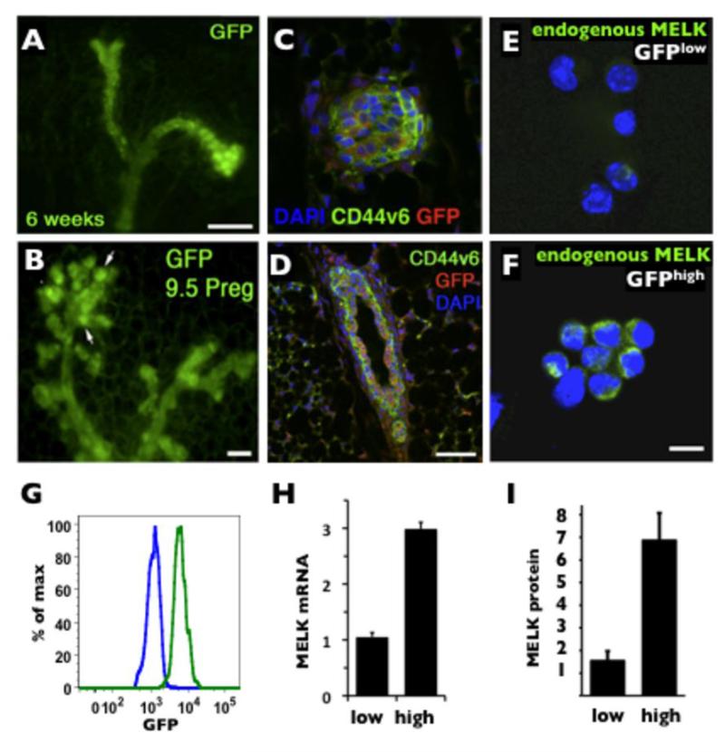
Figure 1. Characterization of MELK expression in normal mammary glands.
A, whole mount mammary fat pad from a 6 weeks-old virgin mouse (Scale bar 100 μm). B, 9.5 days pregnant MELK-GFP female. Note the increased GFP expression in the proliferative alveoli (arrows). Scale bar 50μm. C and D, immunofluorescence of a 6 weeks-old virgin MELK-GFP transgenic mouse mammary gland demonstrating colocalization of GFP expression (red) and CD44v6 (green) in a terminal end bud (C) and predominant GFP expression in epithelial cells as demarcated by CD44v6 in a cross section of a mammary duct (D). Scale bar 30μm. E and F, detection of endogenous MELK protein by immunofluorescence in FACS isolated GFPlow (bottom 10%) and GFPhigh (top 10%) cells. Scale bar 10μm. G. Fluorescent histograms of GFPlow (blue line, median=1.1×10³) and GFPhigh (green line median=5.5×10³) populations. H. Q-PCR detection of MELK mRNA in GFPlow and GFPhigh populations (normalized to 18s expression). I. Quantification of endogenous MELK protein shown in E, F normalized to DAPI).

