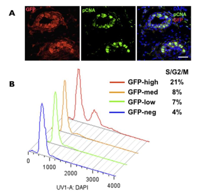
Figure 2. Levels of MELK expression correlate with proliferation in normal mammary cells.
A, colocalization of PCNA (green) and GFP (red) in the luminal cells of the ducts in 6 weeks-old virgin mammary. DAPI staining is in blue. Scale bar 40μm. B, the cell cycle analysis of various GFP populations from freshly dissociated 6 weeks-old virgin mammary glands.

