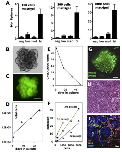
Figure 5. The GFPhigh population in MMTV-Wnt1/MELK-GFP bitransgenic tumors is enriched for tumor initiating cells.
A, GFPHigh population is greatly enriched in spheroid forming activity using a sphere forming culture assay under all conditions tested; p<0.05 for all GFPhigh, one-way ANOVA. Proliferating tumorspheres (B, bright field) retain GFP expression (C). Scale bar 100μm. The matrigel cultures of GFPhigh cells have an increased total number of cells (D) but a decreased frequency of sphere forming cells (E). The mammosphere assay is linear for all cell densities and at all passages tested (F). Colony formation efficiency decreased with the first three passages. 1st passage, red, linear regression y = 0.02x + 4□R2 = 1; 2nd passage, green, y = 0.0138x - 0.4401□R2 = 0.9962; 3d passage, blue, y = 0.0084x - 1.9418□R2 = 0.9385). G. Whole-mount GFP fluorescence of a 20 days outgrowth after transplantation of 10 GFPhigh cells derived from WMG49 primary tumor. Scale bar 1mm. H&E (H) and K8, K14 and DAPI (I) staining of a solid tumor developed from transplanted GFPhigh cells clonally derived from WMG300 primary tumor. Scale bar 50μm.

