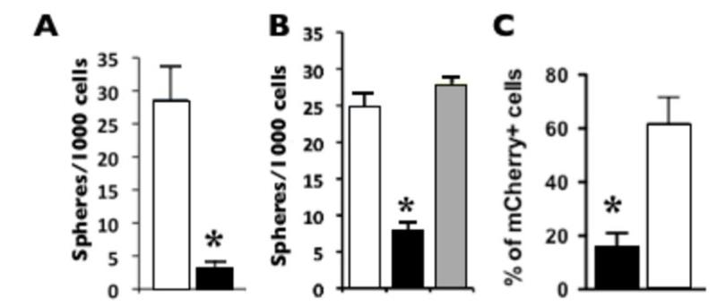
Figure 6. MELK function is required for Wnt1-induced tumorigenesis in vitro and in vivo.
Black bars = MELK shRNA, white bars = control shRNA, grey bar = MELK cDNA rescue. A and B, MELK shRNA reduced the frequency of tumorsphere-initiating cells in Wnt1 tumors. B, co-infection with virus expressing the MELK cDNA rescues tumorsphere proliferation; p < 0.05, n = 3. C, the percentage (%) of mCherry-positive cells in tumors (from D) after transduction with MELK shRNA is reduced compared to control shRNA. Unpaired t-test, p=0.0072.

