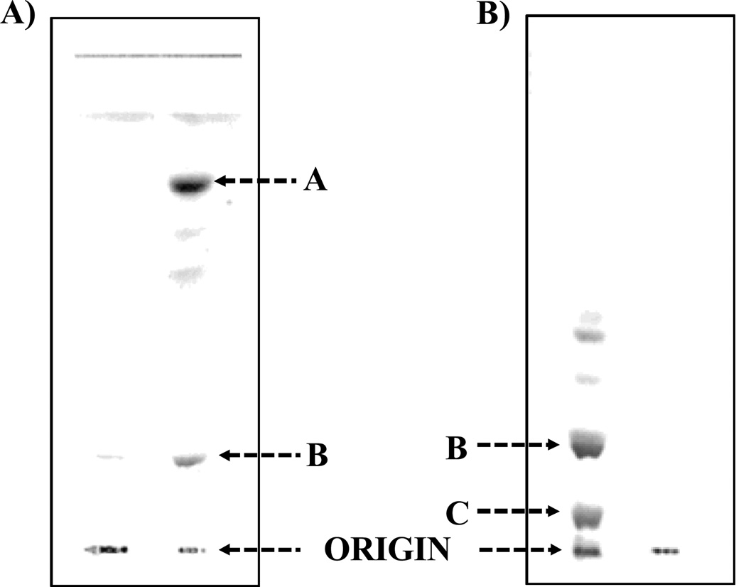
Figure 1.
Thin layer chromatography (TLC) was performed on silica gel GHL plates with a solvent system composed of chloroform/methanol/water/ammonia (50:25:4:2, v/v/v/v). 1A illustrates the TLC analysis of the aqueous phase purified LPS of F. tularensis (List Biologicals, 20 µg, Lane 1, origin) and lipid A bound to LPS of F. tularensis (Lane 2). Band A (Lane 2) contains the unphosphorylated lipid A species and band B (Lane 2) represents the phosphoryl galactosamine lipid A species. 1B illustrates the TLC analysis of the F. tularensis LVS (CDC) LPS (Inzana group) prepared before and after the chloroform/methanol (2:1,v/v) extraction. The free lipid A fractionated into this extract and the LPS was purified away from it. Lane 1 is the free lipid A species and LPS (origin) and 2 is the purified LPS. Band B is composed of the phosphoryl galactosamine lipid A species and was present predominantly in the aqueous layer. In contrast, band C contains the phosphorylated lipid A species with both a galactosamine and hexose residue and was only present in the phenol layer.

