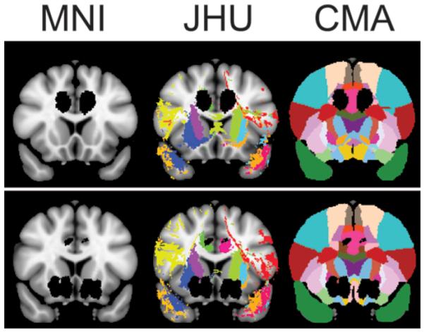
Figure 4.

Representative lesion mask projected on coronal T1-weighted Montreal Neurological Institute (MNI) MNI152 template, Johns Hopkins University (JHU) white-matter tractography atlas, and Center for Morphometric Analysis (CMA) structural atlas. Lesion masks are shown in black, and each color represents a unique atlas region of interest. Upper: Anterior cingulotomy. Lower: Limbic leucotomy.
