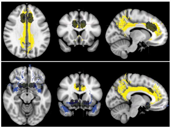
Figure 6.
Representative lesion masks with selected Johns Hopkins University white-matter tractography atlas ROIs. Lesions are shown in black. Selected ROIs are the cingulum (yellow) and uncinate fasciculus (blue). Lesions and ROIs are projected on the T1-weighted MNI152 template. Upper: Anterior cingulotomy. Axial, coronal, and sagittal images (left to right). Lower: Limbic leucotomy. Axial, coronal, and sagittal images (left to right).

