
Figure 3. Nrf2 activation enhances the degradation of phosphorylated tau in neurons.
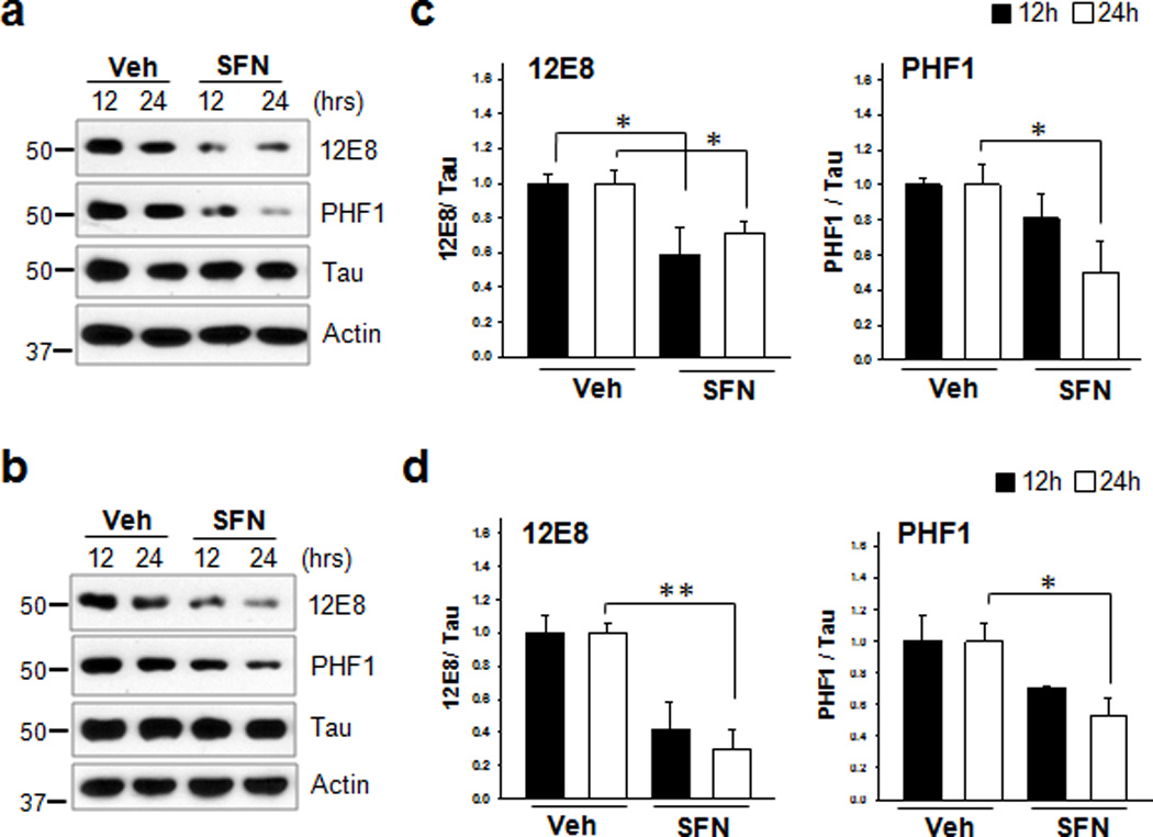
(a) Primary cortical neurons (DIV 5) were treated with sulforaphane (10 µM, SFN) for 12 h or 24 h. (b) Stably transfected CN1.4 cortical neurons were maintained in the presence of doxycycline (1 µg/ml) to induce the expression of tau for 24 h, and subsequently treated with SFN for 12 h or 24 h. The levels of tau phosphorylated at Ser262/Ser356 and Ser396/Ser404 were analyzed by immunoblotting using the 12E8- and PHF1 antibodies, respectively. Tau was detected with a polyclonal phospho-independent tau antibody (Tau). The relative molecular masses (kD) are indicated to the left of each blot. (c, d) Bar graphs represent the relative optical density of phosphorylated tau normalized with that of total tau. Data shown are mean±SE of three independent experiments and were analyzed using Student’s t test. (*, p<0.05; **, p<0.01)