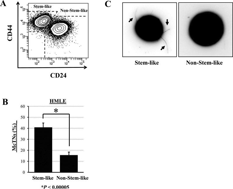
Figure 1. Stem-like mammary epithelial cells have increased microtentacles.
A) HMLE cells have distinct subpopulations of stem-like (CD44hi/CD24lo) and non-stem-like (CD44lo/CD24hi) cells. B) Flow sorted stem-like subpopulations of HMLEs display significantly higher microtentacle frequencies than non-stem-like subpopulations. Columns, mean for three blinded experiments where at least 100 CellMask-stained cells were counted, representative of three independent experiments; bars, SD (P ≤ 0.00005, t test, black asterisk). C) Phase-contrast images of detached HMLE subpopulations where stem-like HMLEs display increased microtentacles (black arrows) compared to non-stem-like HMLEs.

