Figure 1.
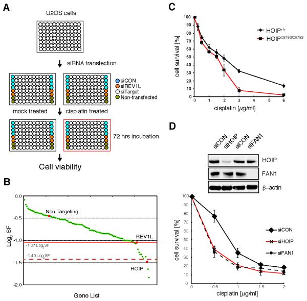
siRNA screen identifies HOIP as an enhancer of cisplatin-induced genotoxicity. A, schematic illustration of the primary siRNA screen, using the siRNA “ubiquitome” library in U2OS cells. B, maximum log2SF is plotted for each gene that was classed as cisplatin-hypersensitive in the primary screen. C, paired wild-type HOIP+/+ and HOIPC879S/C879S knock-in mouse embryonic fibroblasts; cell viability was assayed 72 hrs after cisplatin treatment by MTS cell proliferation assay. D, HEK293 cells were transfected with the indicated siRNAs and cell lysates were assayed for efficient knockdown by immunoblotting with the indicated antibodies (top panel). Cell viability was assayed as in (C). Data in (C) and (D) are represented as mean ± SEM from three independent experiments.

