Figure 3.
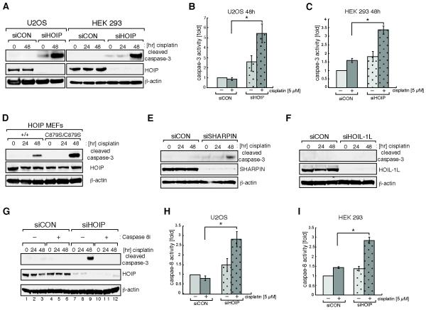
HOIP depletion sensitizes cells to cisplatin-induced apoptotic cell death. A, U2OS (left panel) or HEK293 (right panel) cells were transfected with mock siRNA (siCON) or siRNA targeting HOIP (siHOIP) and treated with 5 μM cisplatin for the indicated time. Cell lysates were analyzed by immunoblotting with the antibodies indicated. B, siCON or siHOIP transfected U2OS cells were treated with 5 μM cisplatin for 48 hours before caspase-3 activity was measured using a caspase-3 Glo assay; * indicates a p-value of 0.0028 C, HEK293 cells were treated as described in (B); * indicates a p-value of 0.0222. D, paired wild-type HOIP+/+ and HOIPC879S/C879S knock-in mouse embryonic fibroblasts were treated with 3 μM cisplatin for the indicated time. Cell lysates were analyzed by immunoblotting. E and F, as (A) except that cells were transfected with siSHARPIN or siHOIL-1L. G, siCON or siHOIP transfected HEK293 cells were treated with vehicle only (−) or 20 μM Z-IETD-FMK caspase-8 inhibitor (+) one hour before treatment with 5 μM cisplatin for the indicated times. Cell lysates were analyzed by immunoblotting. H, as (B) except that a caspase-8 Glo assay was used to measure caspase-8 activity; * indicates a p-value of 0.0076. I, HEK293 cells were treated as described in (H). * indicates a p-value of 0.014. Data in (B), (C), (H) and (I) are represented as mean ± SEM from three independent experiments and Student’s t-test was used to calculate significance.

