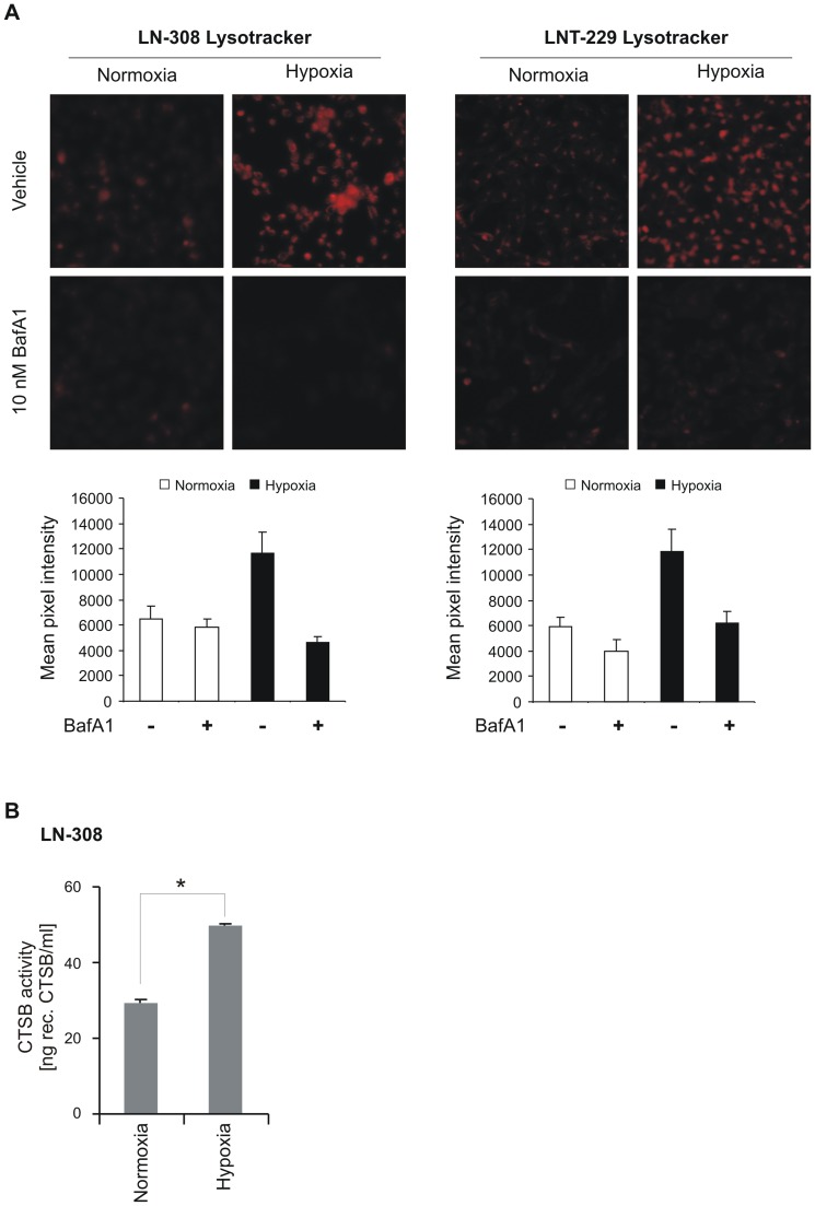
Figure 3. Hypoxia increases lysosomal acidification and cathepsin B activity.
A, LN-308 or LNT-229 cells were grown under normoxia or hypoxia (0.1% O2) for 10 h and thereafter the lysosomal compartment was stained using Lysotracker red. Representative images are shown. Mean pixel intensities were measured with ImageJ software (n = 6, SD, *p<0.05). B, cathepsin B activity in LN-308 cells was measured after 8 h of hypoxia.

