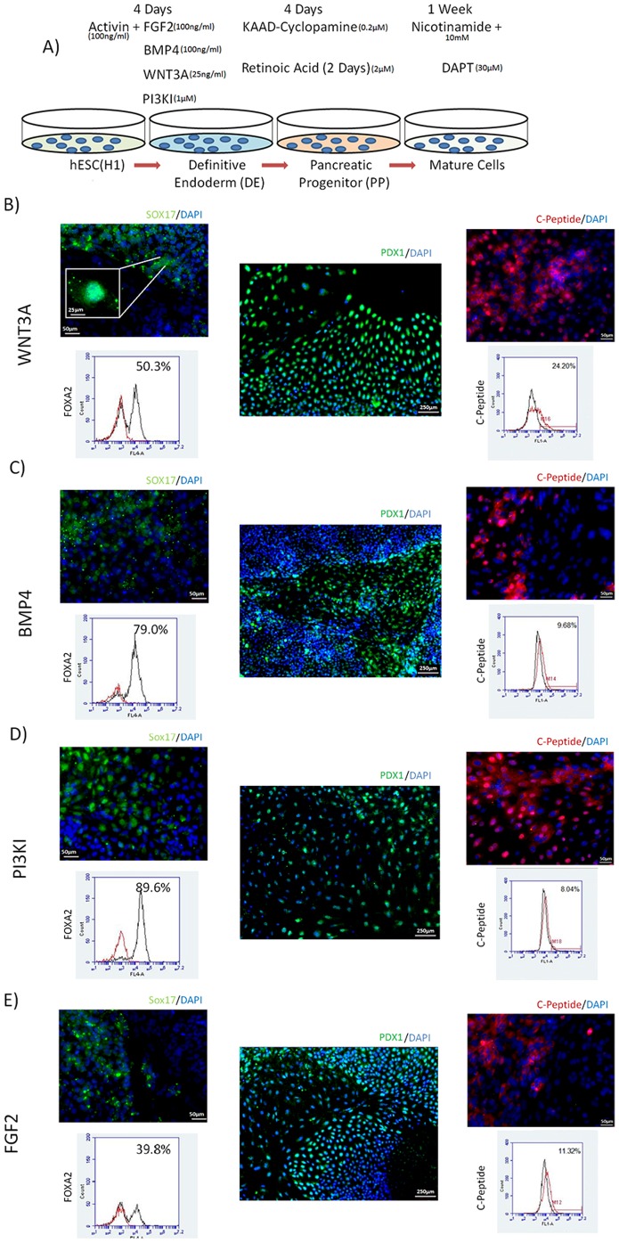
Figure 1. Multi-stage Differentiation System.
(A) Schematic representation of multi-stage differentiation system. Detailed media formulation found in Supp table 1. DE was induced by modulation of nodal pathway simultaneously with one of four alternate pathways. PP was achieved by SHH inhibition along with retinol signaling. Maturation was induced by notch inhibition. Differentiation using WNT3A (B), BMP4 (C), PI3KI (D) or FGF2 (E) at DE stage. IF pictures show nuclear staining of SOX17 (green) and Flow cytometry shows yield of FOXA2 after DE induction, followed by nuclear PDX1 IF pictures (purple) after PP induction and cytoplasmic C-Peptide IF (red) expression yield as measured by flow cytometry after maturation.

