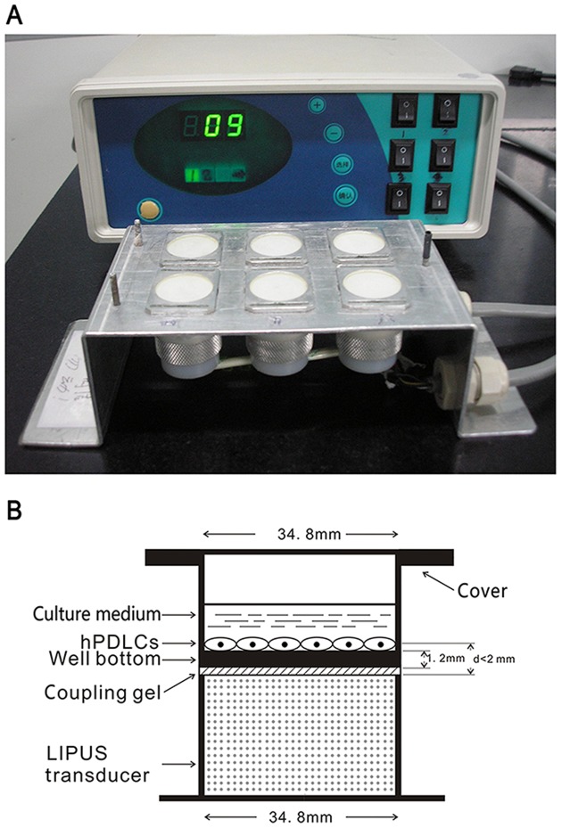
Figure 1. Schematic illustration of ultrasound device and procedure.
(A) Schematic view of the LIPUS exposure device used in this study, which consists of an array of 6 transducers. (B) Schematic illustration showing that a culture plate is placed on the ultrasound transducer array with a thin layer of ultrasonic coupling gel, and the distance between the transducer and cells was less than 2 mm.

