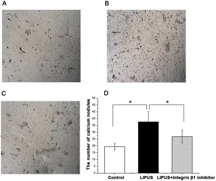
Figure 9. Detection of calcium nodules by Alizarin red staining in hPDLCs.
HPDLCs were assigned into LIPUS-treated group, non-treated control group and the combination group treated wtih LIPUS and integrinβ1 inhibitor, cultured with the osteoblast inducing conditional media for up to 21 days. The mineralized matrix deposition was determined using alizarin red staining. (A) The formation of calcium nodules in non-treated control cells. (B) The formation of calcium nodules in LIPUS-treated cells. (C) The formation of calcium nodules in cells treated wtih both LIPUS and integrinβ1 inhibitor. (D) LIPUS stimulation promoted formation of calcium deposition in hPDLCs, while a significant decline when the specific integrinβ1 inhibitor was used. The data are presented as the mean±SD of three separate experiments. * P<0.05 vs. any of the other two groups.

