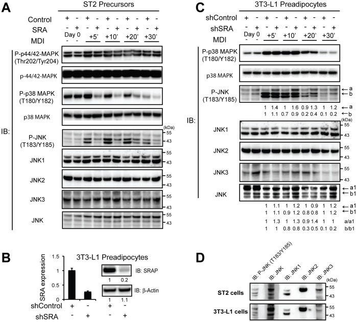
Figure 3. SRA regulates p38/JNK activity during early preadipocyte differentiation.
A, Control or SRA-overexpressing ST2 cells were grown to confluence and induced to differentiate with MDI. Cell lysates were obtained at the indicated times post-MDI induction. Phosphorylation and total protein expression of p38, p44/42 and JNK was assessed by immunoblotting using specific antibodies, as indicated. B, 3T3-L1 preadipocytes with stable knockdown of SRA were generated by retroviral infection with an shRNA against SRA (shSRA); a scrambled shRNA was used to generate control preadipocytes (shControl). Left panel, stable knockdown of endogenous SRA RNA in 3T3-L1 preadipocytes was determined by RT-qPCR using mouse SRA primers. Transcript expression was normalized to Ppia (cyclophilin A) and is presented as mean ± S.D. relative to expression in shControl cells set at 1. Right panel, immunoblot using an SRAP specific antibody confirmed the effective knockdown of endogenous SRA protein (SRAP) in shSRA 3T3-L1 preadipocytes. Reprobing with anti-β-actin served as a loading control. Bands were quantified from immunoblot digital images using Bio-Rad Quantity One software, and the relative results are presented below each immunoblot image. C, shSRA and shControl 3T3-L1 preadipocytes were induced with MDI and protein phosphorylation assessed as described for cells in Figure 3A. Bands labeled a and b in the P-JNK immunoblot, and a1 and b1 in the total JNK immunoblot, correspond to the p54 and p46 kDa species and were quantified as stated above. Results in A, B and C are representative of three independent experiments. D, For ST2 cells, the sample +10 minutes MDI minus SRA was loaded into multiple lanes of one gel and immunoblotted. The membrane was cut so that each lane could be probed individually with an antibody to either phosphoJNK (P-JNK), total JNK (JNK), JNK1, JNK2 or JNK3. The lanes were reassembled to capture the digital image shown. A similar procedure was used for 3T3-L1 cells +10 minutes MDI +shSRA.

