
Figure 4. Depletion of SRA decreases autophosphorylation of insulin receptor β, tyrosine phosphorylation of IRS-1 and expression of IRβ.
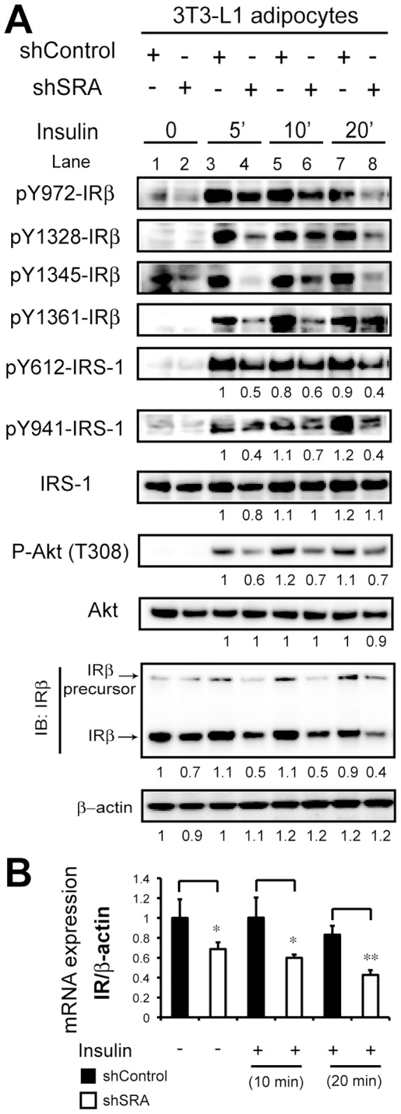
3T3-L1 preadipocytes with stable knockdown of SRA (shSRA) or with shControl infection were induced to differentiate with MDIT. At day 11 post-induction, adipocytes were serum-starved for 12 h followed by treatment with or without insulin (10 nM) for the indicated durations. A, Total cell lysates were subjected to immunoblotting (IB) with antibodies against the indicated phospho-proteins or total IRβ, IRS-1, or Akt; β-actin was used as a loading control. Expression of pY612 and pY941-IRS-1, IRS-1, P-Akt, Akt, IRβ and β-actin was quantified from immunoblot digital images using Bio-Rad Quantity One software. As indicated below the IRβ blotting, expression of P-Akt and IRβ protein was normalized to expression of either Akt or β-actin and the signal of each band was compared to that for control cells as indicated (for which the level of expression was set at 1). B, Gene expression of IR mRNA at each condition was analyzed by RT-qPCR and was normalized to the expression of β-actin.