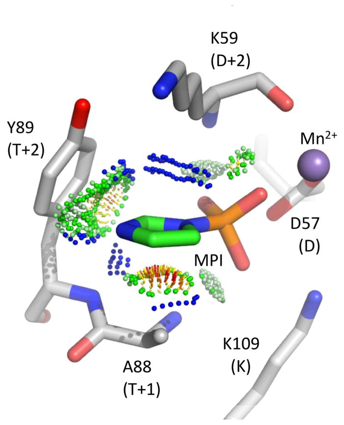
Figure 3.
Model of MPI docked into the active site of CheY *KY·Mn2+· BeF3− (PDBid 3FFW)25. Green and blue probe dots represent favorable van der Waals interactions whereas orange and red dots represent steric clashes. The BeF3− ion present in the original structure is not shown for clarity.

