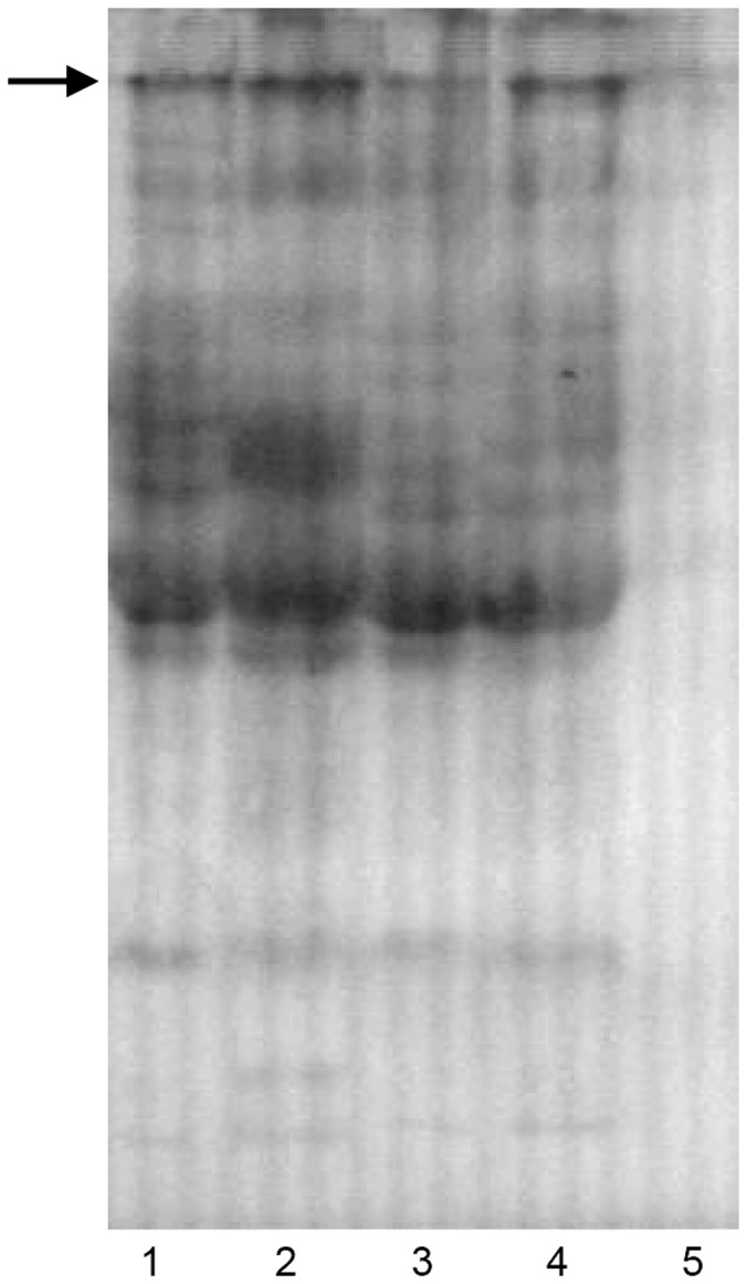
Figure 2. Western blot of plasma from treated and control male fish.

Lane 1: plasma from E2 injected fish (20×); Lane 2: plasma from effluent exposed fish; Lane 3: plasma from 0.5% diluted MBR outlet exposed fish; Lane 4: plasma from 0.5% diluted leachate exposed fish; Lane 5: plasma from negative control male. Primary antibody: rabbit anti-goldfish Vtg serum (diluted 1∶600). Secondary antibody: goat anti-rabbit IgG horseradish peroxidase conjugate (diluted 1∶1600).
