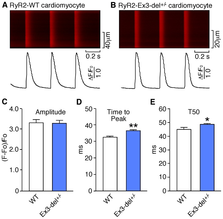
Figure 4. Depolarization-induced Ca2+ transients in WT and heterozygous RyR2 Ex3-del mutant cardiomyocytes.
Ventricular myocytes isolated from RyR2 WT and Ex3-del+/ − mutant hearts were loaded with Rhod-2-AM and perfused with 2 mM extracellular Ca2+ in KRH solution and paced at 3Hz. Ca2+ transients were monitored by line-scan confocal Ca2+ imaging. Representative images/traces of WT (A) and Ex3-del+/ − mutant (B) cardiomyocytes, and average data of the amplitude (C), time to peak (D), and time to 50% decay (E) of Ca2+ transients in WT and Ex3-del+/ − mutant cells are shown. Data shown are mean ± SEM from 35 WT and 58 mutant cells (**P<0.001; *P<0.05).

