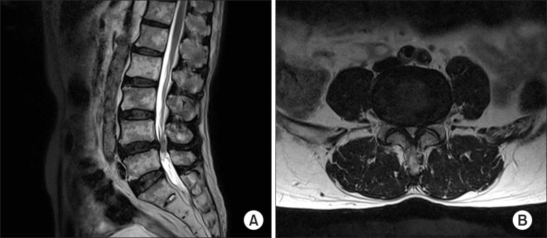
Fig. 2.
T2-weighted magnetic resonance images (MRI) of the lumbar spine in a 75-year-old man suffering from pain in his back, both thighs, and calves that persisted for seven months. (A) Sagittal and (B) axial views of the MRI show central stenosis at the L4-5 level caused by a bulging disc, facet arthrosis, and thickening of the ligamentum flavum.

