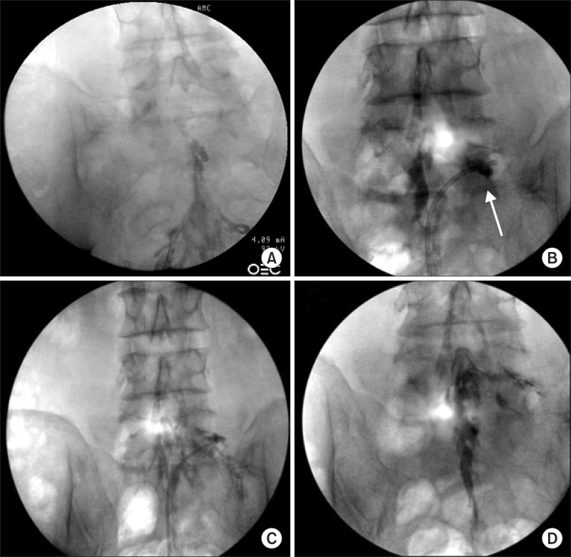
Fig. 3.
Serial fluoroscopic images of percutaneous epidural adhesiolysis (PEA) using the inflatable balloon neuroplasty catheter. (A) Anteroposterior view verified before the procedure showing filling defects of contrast medium at the epidural space above the level of L5-S1 and both L5 intervertebral foramina. (B) Fluoroscopic view showing the inflatable balloon neuroplasty catheter placed in the L5 intervertebral foramen and the balloon filled with contrast medium. Foraminal stenosis is visualized by the degree of distortion of the balloon (arrow). (C) Decompression is performed along the intervertebral foramen by ballooning. (D) After decompression along the pass from the lateral recess to the intervertebral foramen, the contrast agent spread well. In addition, the contrast agent in the epidural space spread upward above the level of L5-S1.

