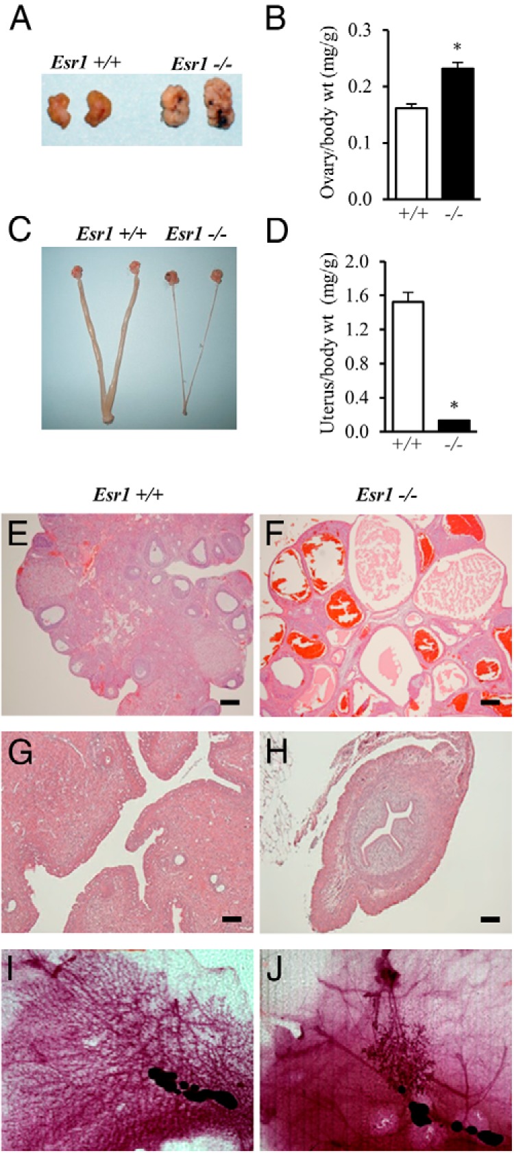
Figure 5.

Effects of ESR1 disruption on the female reproductive system. A–D, Reproductive tracts of adult wild-type and Esr1-null females (8 weeks of age) were examined, including gross appearance and weights for ovaries (A and B) and uteri (C and D). Sample sizes for the organ weight measurements were ≥10 per genotype. Asterisks indicate a significant difference between the genotypes: *, P < .001. E–H, Representative hematoxylin- and eosin-stained tissue sections from ovaries (E and F) and uteri (G and H) from wild-type and Esr1-null females. I and J, Whole-mount staining of mammary glands from wild-type (I) and Esr1-null (J) females. Please note the cystic and hypoplastic features of ovaries and uteri, respectively, and the rudimentary ductal and alveolar structures in the mammary glands of Esr1-null females (F, H, and J). Scale bars, 0.25 mm.
