Figure 3.
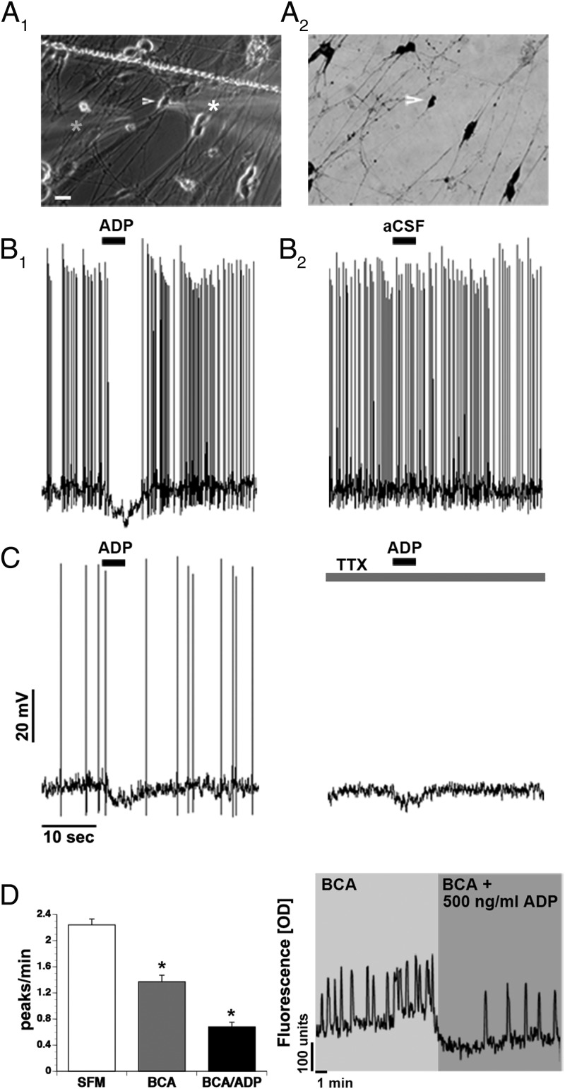
ADP inhibits GnRH neurons directly. A, Whole-cell recordings of GnRH neurons (A1 arrow/white star) showed an acute response to 5-μg/mL ADP applied with a pipette (A1 gray star). Explants were fixed and stained for GnRH after recordings were completed (A2). B, Example of spontaneously active GnRH neuron inhibited by a 4-second application of ADP (B1), replacing the ADP in the electrode with aCSF (B2). C, Example of GnRH neuron inhibited by a 4-second application of ADP ± TTX. D, The combined application of BIC/CNQX/AP5 (BAC) decreased the frequency of calcium oscillations in GnRH neurons. Coapplication with ADP (500 ng/mL) further decreased the neuronal activity. A representative calcium imaging recording showing spontaneous baseline oscillations in intracellular calcium levels of a GnRH neuron during 10-minute periods of BAC and BAC/ADP. Line in A1, scratch in coverslip. Scale bar: 10μM (A1 and A2).

