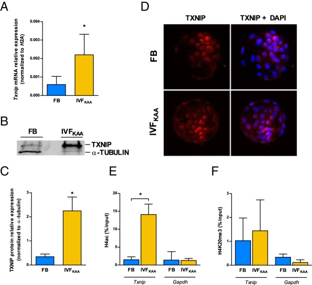
Figure 5.
Misregulation of Txnip in IVFKAA blastocysts. A, Txnip mRNA expression relative to H2A in FB (blue) and IVFKAA (yellow) embryos. B, Western blot of TXNIP and β-actin protein with (C) quantification of TXNIP relative expression for both conditions. D, Immunostaining of TXNIP localization (red) with 4′,6-diamidino-2-phenylindole (DAPI) counterstain (blue). E and F, Chromatin immunoprecipitation of (E) H4 acetylation and (F) H4K20me3 enrichment at the Txnip and Gapdh promoters in FB and IVFKAA blastocysts. Error bars depict SD. *, P < .05.

