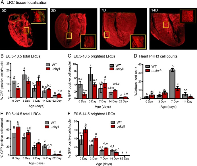
Figure 3.
Cardiac LRCs. A, 3-dimensional confocal images of whole hearts (×50) constructed using Metamorph software. Hearts were sampled from 0-, 3-, 7-, and 14-day-old pups (0 day old, 0D etc; red, phalloidin-stained actin; green, LRCs) pulsed with doxycycline from E0.5–E10.5. Inset images (×250) correspond to areas within yellow boxes. B and C, The number of total and brightest LRCs was normalized to total nuclei and quantified in tissues sampled on the indicated days. D, The number of GFP-positive cells also positive for PHH3 was quantified in tissues sampled on the indicated days (n = 3/group). E and F, Total and brightest LRCs in mice pulsed with doxycycline from E0.5–E14.5. In all graphs, different letters indicate statistical significance (P ≤ .05, n = 5–7/group) between any particular group whereas the same letters indicate no differences.

