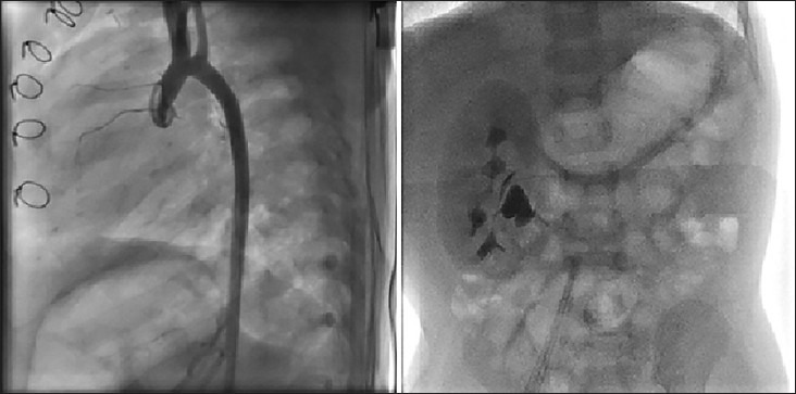
Figure 1.

Left panel: Shows the aortogram in lateral view in a 1-year-old male infant after surgical correction for interrupted aortic arch and large ventricular septal defect at the age of 6 months. The abdominal fluoroscopy at the end of the procedure showed lack of any functioning left kidney. Right panel: Renal ultrsonography shows left non-functioning multicystic cystic kidney disease. This patient had normal blood urea nitrogen and creatinine both before and 24 h after the cardiac catheterization and angiography
