Figure 6.
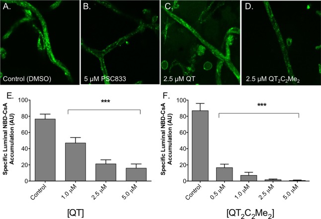
P-gp-mediated transport in isolated rat brain capillaries. P-gp transport activity was determined using isolated rat brain capillaries as described in Methods. Representative images of capillaries preincubated with (A) DMSO, (B) 5 μM PSC833, (C) 2.5 μM QT, or (D) 2.5 μM QT2C2Me2 (8) followed by 60 min incubation with 2 μM NBD-CsA. Capillary fluorescence was visualized with a Zeiss 510 NLO confocal scanning microscope and quantitated from images using ImageJ software. (E, F) Capillaries were preincubated with the indicated concentrations of (D) QT and (E) QT2C2Me2 (8) followed by 60 min incubation with 2 μM NBD-CsA. Shown are mean ± SEM for 5–12 capillaries. Statistical comparisons: one-way ANOVA, ***P < 0.001.

