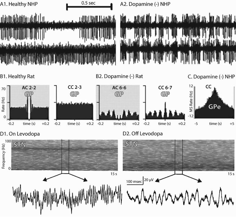
Fig. 2.
Electrophysiological phenomena observed in parkinsonian subjects. Single unit pallidal recordings from dopamine-intact (A1) and MPTP-treated (A2) non-human primates (NHP). The upper trace in A1 shows a typical GPe “high-frequency discharge with pauses” pattern. Pallidal burst-firing is increased after MPTP treatment [104]. (B) Auto- and cross-correlograms of single unit activity from anesthetized rats during “cortical activation,” emphasizing the emergence of synchronized beta oscillations. AC–autocorrelogram, CC–cross-correlogram. Numbers identify individual units (for example CC 2-3 is the cross-correlogram between units 2 and 3 [92]). (C) Cross-correlogram illustrating non-oscillatory synchrony among GPe neurons in an MPTP-treated NHP [59]. The ordinate displays the mean-subtracted (MS) firing rate. (D) Time-frequency plots and sample LFPs from the STN of a patient with PD on (D1) and off (D2) levodopa [89]. (Figures adapted with permission from the relevant sources).

