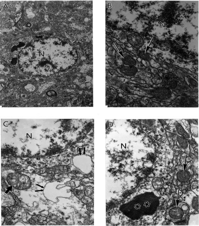
Figure 4.
The ultrastructural characteristics of the neuronal mitochondria from AD brain biopsy. A. Neurons with different degrees of ultrastructural lesions. Partially and completely damaged mitochondria are mostly located in the neuronal cell body and coexist with lipofuscin formation. Original magnification X 5,000. B. Large numbers of electron-dense hypoxic mitochondria (indicated by single arrows) were present throughout the cell body and characterized the abnormal mitochondrial cristae. Original magnification: X 20,000. C. Partially (indicated by single arrow) and completely damaged (double arrow) mitochondria. Original magnification X 20,000. D. The neuronal cell body shows the presence of hypoxic mitochondria (indicated by single arrows) close to lipofuscin (double asterisk). Original magnification X 20,000. Abbreviations used in figure: N– neuronal nucleus (reprinted from [87] with permission).

