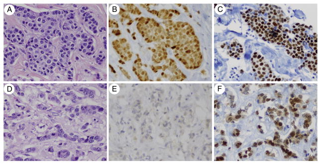
Fig. 2.

GATA3 labeling is seen in MBCs that have loss of ER and/or PR expression from the primary to the metastasis. The primary invasive ductal carcinoma (A) is diffusely positive for ER (B) and GATA3 (C). The paired MBC to the liver harvested at autopsy (D) shows complete loss of nuclear ER labeling (E), but intact and diffuse GATA3 labeling (F). Normal ovarian stroma harvested at autopsy showed positive nuclear ER labeling and served as a positive control for ER labeling in autopsy tissue (not shown) (hematoxylin and eosin, ER immunostain, and GATA3 immunostain; ×400).
