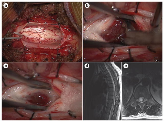
Figure 1.
Surgery and imaging in spinal cord astrocytoma. a–c | Intraoperative images showing the excision of a diffuse intramedullary astrocytoma in the spinal cord from a posterior approach. The spinal cord appears enlarged, and the surgical approach begins with a midline myelotomy to separate the dorsal columns (a). The tumour is then exposed and excised via careful dissection (b). Gliosis and hyperaemia can be seen in the resection cavity (c). d, e | Preoperative sagittal (d) and axial (e) T2-weighted MRI reveal hyperintensity and expansion of the spinal cord.

