
Figure 4.
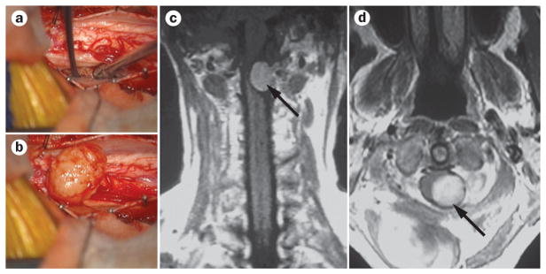
Surgery and imaging of spinal cord meningioma. a, b | Intraoperative images showing pre-exposure (a) and postexposure (b) of a spinal meningioma. c, d | Coronal (c) and axial (d) postgadolinium MRI demonstrating a cervical extramedullary meningioma (arrows) with avid enhancement.