Figure 6.
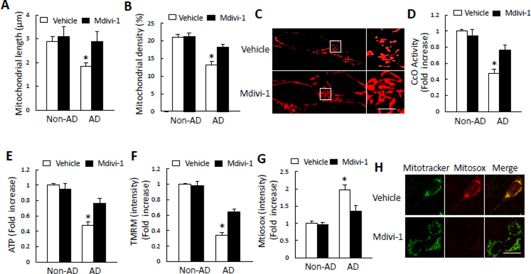
Inhibition of DLP1 by mdivi-1 rescues mitochondrial structure and function in AD cybrid mitochondria. A–C) Effect of mdivi-1 on mitochondrial morphology. Mdivi-1 treatment (10 µM for 24 h) significantly increased mitochondrial length (A) and density (B) Representative images for Mitotracker red staining reveal mitochondrial morphology (C). *p<0.05 versus all other groups. The right panel is a larger image corresponding to the indicated image on the left panel (Scale bar = 5 µm). D–H) Effect of mdivi-1 on mitochondrial function. Treatment with mdiv-1 resulted in increased CcO activity (D), ATP levels (E), and TMRM intensity in AD cybrid cells (F). Mdivi-1 also attenuated mitochondrial ROS production as shown by reduced level of Mitosox staining intensity (G) Representative images for Mitotracker and Mitosox staining are shown (H) (Scale bar = 10 µm). *p<0.05 versus all other groups. N = 5–7 cell lines/group.

