Figure 3.
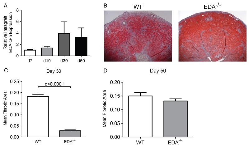
EDA cFN promotes fibrotic remodelling associated with chronic cardiac allograft rejection. (A) Kinetic time course of intra-graft EDA cFN transcript levels in WT recipients transiently depleted of CD4+ cells. Bars represent mean and SEM of three individual grafts. (B) Representative Masson’s trichrome-stained sections of cardiac allografts harvested on day 30 post-transplant. (C) Morphometric quantification of allograft fibrosis on day 30 post-transplant in WT and EDA−/− graft recipients. Bars illustrate mean and SEM of a minimum of ten frames of view from at least eight independent transplants. (D) Morphometric quantification of allograft fibrosis on day 50 post-transplant in WT and EDA−/− graft recipients. Bars illustrate mean and SEM of a minimum of ten frames of view from at least six independent hearts.

