
Figure 1.
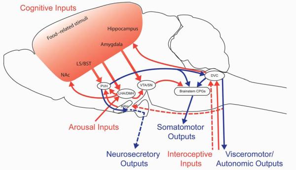
Outline of circuits involved in controlling feeding behavior. The diagram depicts a subset of the neuronal connections reported to influence feeding, chosen to highlight opportunities for integration between different classes of inputs (red arrows) and outputs (blue arrows). These inputs and outputs are transmitted via neuronal projections (solid lines) as well as humoral factors (dotted lines). Note that arrows reflect connectivity between two brain regions and not an actual anatomical pathway. Interoceptive inputs transmit information about energy availability that is essential for homeostatic regulation of food intake. Sensory meal-related signals from the alimentary tract are relayed to the brain through autonomic and sensory nerves, as well as humoral factors. Cognitive inputs are required for volitional feeding; food-related cues are processed in cortico-limbic circuits to confer learned aspects of feeding as well as to determine the reward value of food. A state of arousal is transmitted by neurons in the LHA to a network of highly interconnected neurons in the hypothalamus and brainstem. Feeding behavior is directed through the coordinated signals of three classes of output pathways. Somatomotor outputs from brain stem CPGs direct the muscular contractions needed to seek and consume food. Visceromotor outputs through the autonomic nervous system regulate the secretion of factors involved in nutrient processing, metabolism and storage. Neurosecretory outputs regulate the release of pituitary hormones that influence feeding behavior. Abbreviations: ARH, arcuate nucleus of the hypothalamus; BST, bed nuclei of the stria terminalis; CPG, central pattern generator; DMH, dorsomedial nucleus of the hypothalamus; DVC dorsovagal complex (area postrema, nucleus of the solitary tract, and dorsal motor nucleus of the vagus); LHA, lateral hypothalamic area; LS, lateral septum; NAc, nucleus accumbens; PVH, paraventricular nucleus of the hypothalamus; SN, substantia nigra; VTA, ventral tegmental area.