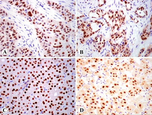
Fig. 2.
GATA3 expression in adult tissue. A. Epidermal cells are positive. Note also positive dermal lymphocytes. B. Luminal cells of ducts of a breast lobule a positive, whereas myoepithelial cells are negative. C. Ureteral epithelium and some stromal lymphocytes are positive. D. Distal tubular epithelial and mesangial cells are GATA3-positive.

