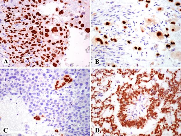
Fig. 4.
A. Examples of GATA3-positive epithelial neoplasms. Strong expression in cutaneous squamous cell carcinoma B. Pancreatic ductal adenocarcinoma cells show nearly uniform labeling. C. Renal chromophobe carcinoma cells are positive. D. Most cells of renal oncocytoma are positive and there is also weak cytoplasmic labeling.

