
Figure 4.
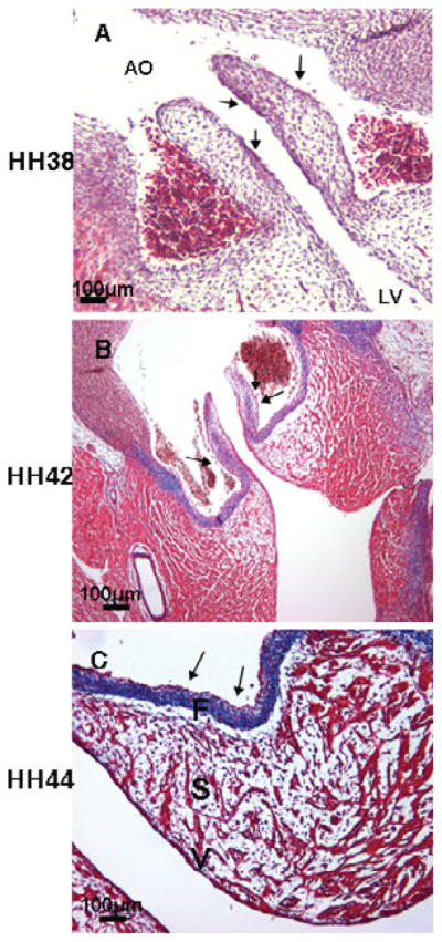
Collagen fiber staining in developing chick embryonic semilunar valves by Masson’s trichrome. Collagen staining was detectable in the fibrosa and ventricularis at HH 38 (arrows, A). By HH 42, collagen fiber staining was constrained in the distal part of the semilunar leaflets and the fibrosa (arrows, B). Condensed collagen fiber staining was observed only in the fibrosa at HH 44 (arrows, C). AO: aorta; F: fibrosa; S: spongiosa; V: ventricularis. Scale bar: 100 μm.