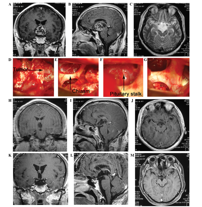
Figure 2.
Case of a 45-year-old male who presented with general fatigue for two months, polyuria with blurred vision and memory loss for >1 month. The patient was diagnosed with a CP. During surgery, the tumor was found to originate from the pituitary stalk. In order to completely remove the tumor, the pituitary stalk was also removed. Three years following surgery, the patient resumed work and there was no evidence of recurrence in MRI scans. (A) The coronary view of CE MRI showing that the tumor was located in the sellar and parasellar area. The lesion is mainly solid with contrast enhanced walls. The borders of the tumor are distinct with absence of peritumoral edema. Mild compression of the right side of the third ventricle can also be noted. It also demonstrates the pituitary stalk, the optic nerve and optic chiasm (see arrow). (B) The sagittal view of CE MRI and (C) axial view, shows the relationship between the tumor and the clinoid part of the ICA, optic nerve as well as optic chiasm. (D, E, F and G) The intra-operative findings of the lesion: the tumor was located retrochiasmatically and below the clinoid part of ICA. The pituitary stalk was invaded by the tumor (see arrow). (H, I and J) Post operative CE MRI depicts complete excision of tumor together with absence of hemorrhage. (K, L and M) There was no evidence for tumor recurrence in the 1 year follow up. CP, craniopharyngioma; MRI, magnetic resonance imaging.

