
Figure 2.
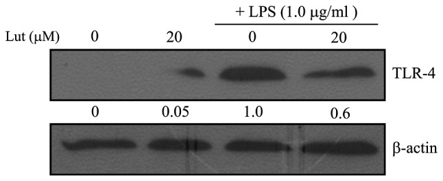
Effects of luteolin on the expression of TLR-4 in LPS-stimulated BV2 microglia. Western blot analysis of TLR-4. Cells were incubated with 20 μM luteolin for 1 h prior to incubation with LPS (1.0 μg/ml) for 24 h. Cell lysates were then prepared and subjected to western blotting. TLR-4 protein expression was quantified by OD ratio using β-actin as a control. Data were collected from three independent experiments each carried out in triplicate. *Indicates a significant difference (P<0.05) relative to cells treated with LPS in the absence of luteolin. TLR-4, Toll-like receptor-4; LPS, lipopolysaccharide; OD, optical density; Lut, luteolin.